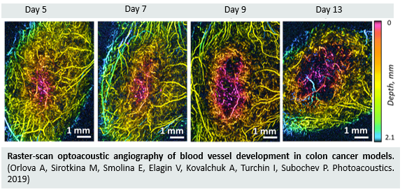
Raster-scan optoacoustic angiography at 532 nm wavelength with 50 μm lateral resolution at 2 mm diagnostic depth was used for quantitative characterization of neoangiogenesis in colon cancer models. Two tumor models of human colon adenocarcinoma (HT-29) and murine colon carcinoma (CT26) different in their histology and vascularization were compared. Tumors of both origins showed an inhomogeneous distribution of areas with high and low vascularization. Rapidly growing CT26 tumor demonstrated a higher rate of vessel growth from the periphery to the center. Peculiarities of the vascularity of tumor models revealed by optoacoustic imaging were confirmed by fluorescent microscopy with FITC-dextran and morphological analysis. The obtained results may be important for the investigation of tumor development and for improvement of colon cancer treatment strategies.
Orlova A., Sirotkina М., Smolina Е., Elagin V., Kovalchuk A., Turchin I., and Subochev P. Raster-scan optoacoustic angiography of blood vessel development in colon cancer models, Photoacoustic 13, 25-32 (2019).
Follow the link below to find out more: