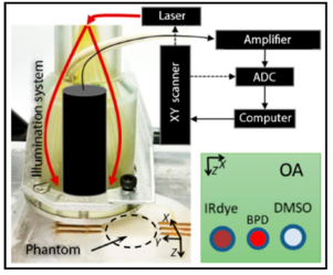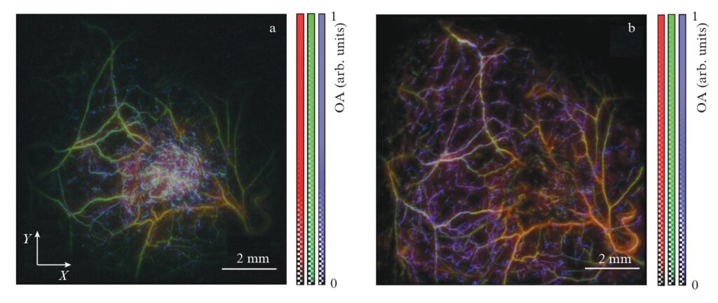
Spherical ultrasonic antennas are used in raster-scan optoacoustic (OA) angiography to record broadband signals generated by haemoglobin molecules in blood when they absorb pulsed optical radiation. Depending on the size of haemoglobin-containing structures, the characteristic frequencies of OA signals can vary quite significantly, ranging from hundreds of kilohertz to hundreds of megahertz. Meanwhile, the bandwidth of the receiving frequency band of standard piezoelectric sensors, as a rule, does not exceed the centre frequency value. It is possible to expand the receiving band of ultrasonic detectors to the required 0.1 kHz – 100 MHz values by using nonresonant piezomaterials based on polyvinidylene fluoride (PVDF). Two ultra-wideband detectors based on PVDF piezofilms of different thicknesses (9 μm and 25 μm) with different amplitude-frequency characteristics are experimentally compared. Comparative OA imaging of a tissue-like phantom demonstrates that the low-frequency sensor (film thickness l = 25 μm) has a greater depth of field, while the high-frequency sensor (l = 9 μm) has a better sensitivity in the range of 40 – 100 MHz. Using OA imaging of an experimental tumour in vivo, it is shown that a sensor with l = 25 μm is better suited for examining normal tissue containing relatively large blood vessels, while a sensor with l = 9 μm is better suited for studying tumour tissue containing a large number of multidirectional blood vessels of minimal size comparable to the maximum spatial resolution of the OA system.
A.A. Kurnikov et al 2021 Quantum Electron. 51 383
Follow the link below to find out more: