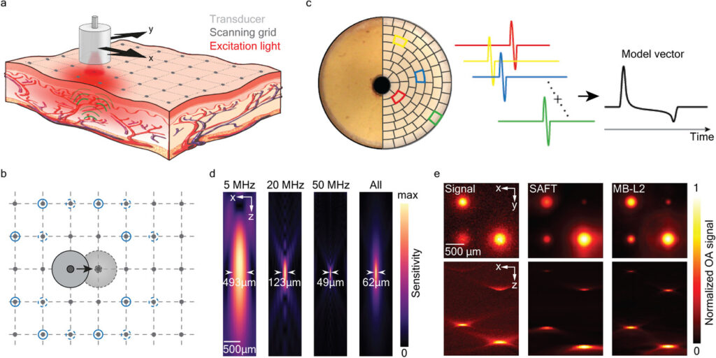
Optoacoustic mesoscopy (OAM) retrieves anatomical and functional contrast in vivo at depths not resolvable with optical microscopy. Recent progress on reconstruction algorithms have further advanced its imaging performance to provide high lateral resolution ultimately limited by acoustic diffraction. In this work, a new broadband model-based OAM (MB-OAM) framework efficiently exploiting scanning symmetries for an enhanced performance is presented. By capitalizing on the large detection bandwidth of a spherical polyvinylidene difluoride film while accurately accounting for its spatial impulse response, the new approach significantly outperforms standard OAM implementations in terms of contrast and resolution, as validated by functional in vivo experiments in mice and human volunteers. Furthermore, L1-norm regularization enables resolving structures separated by less than the theoretical diffraction-limited resolution. This unique label-free angiographic performance demonstrates the general applicability of MB-OAM as a super-resolution deep-tissue imaging method capable of breaking through the limits imposed by acoustic diffraction.
Weiye Li, Urs A. T. Hofmann, Johannes Rebling, Quanyu Zhou, Zhenyue Chen, Ali Ozbek, Yuxiang Gong, Pavel Subochev, Daniel Razansky, Xosé Luís Deán-Ben. Broadband Model-Based Optoacoustic Mesoscopy Enables Deep-Tissue Imaging beyond the Acoustic Diffraction Limit.
Laser Photonics Rev.2022,16, 2100381
Follow the link below to find out more: https://doi.org/10.1002/lpor.202100381