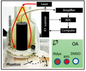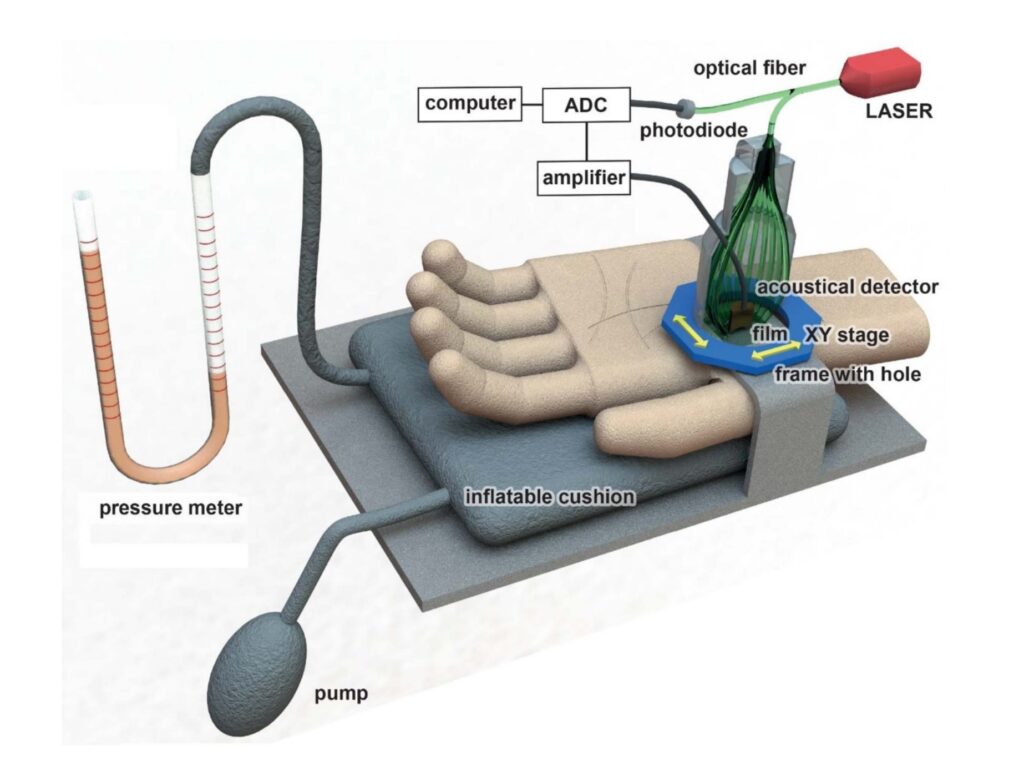
We report on the estimation of blood content and vessel volume fraction changes in the microcirculatory bed of human skin under controlled mechanical compression using 3-dimensional optoacoustic (OA) angiography. A consecutive decrease in the fraction of blood vessels and skin blood content as a result of an increase in pressure from 0 to 72 mmHg applied to the imaged area was demonstrated by means of an acoustical-resolution OA microscope with a spatial resolution of 50 μm. Pressures below 32 mmHg were shown to weakly affect the acquired OA angiograms. The loss of OA signal from the blood vessels was observed after a further pressure increase of up to 72 mmHg. The vascular changes observed by OA microscopy were confirmed by infrared (IR) thermometry measurements which revealed similar dynamics of microcirculation interruption in the area under pressure.
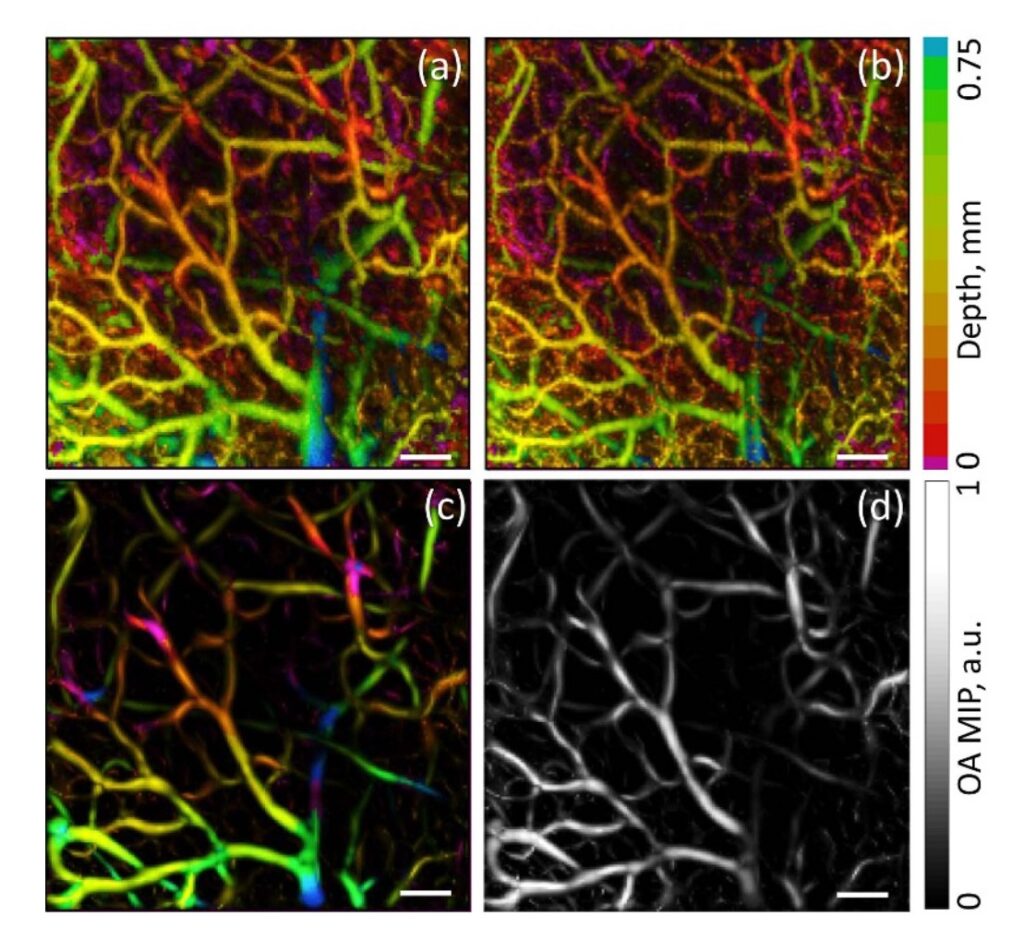
The images represent maximum intensity projections along the depth axis; all bars are 1 mm.
Anosov A. A., Kirillin M. Yu., Orlova A. G., Erofeev A. V., Sharakshane A. S., Shcherbakov M. I., Sergeeva E. A., Saijo Y. and Subochev P. V. Volumetric quantification of skin microcirculation disturbance induced by local compression, Laser Phys. Lett. 17 (2020) 085601 (6pp)
Follow the link below to find out more: