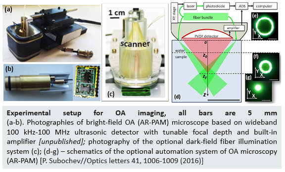
Polyvinylidene difluoride (PVdF) is a piezopolymer with high internal damping. PVdF -based transducers are characterized by ultrawide bandwidth, although typically have a lower sensitivity compared to conventional bulk piezoceramic devices. PVDF transducers are especially useful in imaging applications (including biomedicine), where excellent axial resolution and tight focusing are required.
PVdF ultrasonic transducers are commonly employed in optoacoustic (OA) imaging of biological tissues, also called photoacoustic (PA) imaging.
OA imaging is based on wideband detection of ultrasonic waves generated by biological tissue chromophores absorbing probing optical pulses. Absorbing of optical radiation by hemoglobin molecules induces local thermal expansion within blood vessels, generating an ultrasonic wave. The ultrasonic waves are detected by an ultrasound transducer at the surface of the biological tissue. As a result, OA angiography provides optical contrast and ultrasonic spatial resolution at the ~1cm depths.
Clinical applications of optoacoustic methods of vascular angiography, including vascularization in oncology, subcutaneous melanin-containing neoplasms, atherosclerotic changes in vascular walls, directional biopsy of signal lymph nodes, the reaction of the tumor to therapeutic effects, age-related pathologies in ophthalmology, hemodynamic reactions of the brain.
Acoustic resolution photoacoustic microscopy at the optical wavelength of 532 nm was used to study functional reaction of blood vessels of healthy human skin during cuffed venous occlusion. The high bandwidth of the employed PVdF detector provided the opportunity for raster-scan optoacoustic angiography of both the superficial and deep plexuses at the high resolution of 35/50 microns (axial/lateral). (A. Orlova et. al.//Photoacoustics, 13 2019).
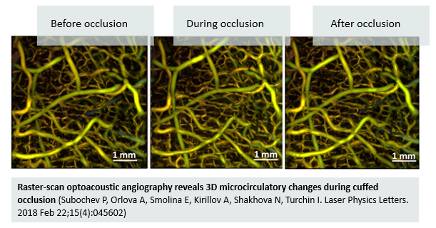
Acoustic resolution photoacoustic microscopy with probing optical wavelength of 532 nm was used to study the functional reaction of blood vessels of healthy human skin during cuffed venous occlusion. The high bandwidth of the PVdF detector provided the opportunity for raster-scan optoacoustic angiography of both the superficial and deep plexuses at the high resolution of 35/50 um (axial/lateral) at ~1mm depths. A reversible increase of blood supply in the microcirculatory bed during occlusion was revealed (P. Subochev et. al. // LPL 15 (4), 2018).